From cell fat to cell fate
How does a cell “decide” what type of cell to become? The question of “cell fate” has been explored for decades now, especially in the context of stem cell biology, but there are still gaps in our understanding. For example, any multicellular organism is made up of different cell types that play specific roles, while they all work together to sustain the organism as a whole.
At the same time, some cell types can transition between different functions. A good example are the skin’s fibroblasts, which form the dermis, between the top layer of epidermis and the bottom layer of fat. Fibroblasts can take on different specializations to help repair wounds, remodel the extracellular matrix, or even cause fibrosis.
This complex system of cell fate has drawn a lot of research, which has mostly focused on external signals from the cell’s microenvironment. By comparison, very little work has been done on possible “internal” processes within the cell that contribute to its specialization.
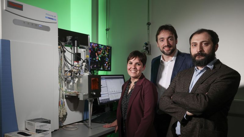
A team of scientists, led by professors Gioele La Manno and Giovanni D’Angelo at EPFL’s School of Life Sciences have now determined for the first time that one of the internal factors to determining a cell’s fate is its production of lipids – fat molecules.
Working on skin fibroblasts, the researchers combined two techniques to sort cells into lipid-producing “profiles”: high resolution mass spectrometry imaging, which allowed them to visualize the distribution of specific lipid inside each cell, and single-cell mRNA sequencing, which allowed them to determine the gene expression profile of each fibroblast – a sort of ID card of what we call a “transcriptome” – and put each cell into a transcriptional subpopulation.
The first thing the study revealed was that dermal fibroblasts can show multiple lipid groups, or “lipid compositional states”, which the researchers dubbed “lipotypes”.
“Cell states are intermediates in the process of cell differentiation where state switches precede terminal commitment,” write the authors.
But there was a clue: each lipotype turned out to correspond to specific transcriptional subpopulations in vitro and to fibroblasts from different skin layers of the skin in vivo.
The question now was what markers could we use to identify the different lipotypes. Given their correlation with the fibroblasts’ transcriptional groups, the researchers proceeded to isolate metabolic pathways that could account for this connection.
They found that the major markers of the different lipid compositional states are a family of fat molecules called “sphingolipids”. Named after the mythical Sphinx, sphingolipids are involved in cell-to-cell communication, as well as protecting the cell’s outer surface by forming barriers on its membrane.
At this point, the researchers made a critical discovery: the different lipotypes influence the different responses that cells have to external stimuli from their microenvironment that “push” them into different cell fates – even if the two original cells were identical. In fact, the researchers found, it is possible to entirely reprogram a cell’s fate by simply manipulating its sphingolipid composition.
In the final leg of the study, the team found that lipid composition and signaling pathways are wired in self-sustained circuits, and it is these circuits that account for the differences between metabolism and gene transcription among fibroblasts.
The key molecule here is fibroblast growth factor, or FGF2, a signaling protein that is involved many processes, such as embryonic development, cell growth, morphogenesis, tissue repair, and even tumor growth and invasion. In the context of this study, sphingolipids were found to regulate FGF2 signaling by using two different types of sphingolipids as positive as negative regulators.
“We uncovered an unexpected relationship between lipidomes and transcriptomes in individual cells,” write the authors, referring to a cell’s complete profile of lipid production. “Lipidome remodeling could work as an early driver in the establishment of cell identity, and following lipid metabolic trajectories of individual cells could have the potential to inform us about key mechanisms of cell fate decision. Thus, this study stimulates new questions about the role of lipids in cell fate decisions and adds a new regulatory component to the self-organization of multicellular systems.”
